This blog post was originally published at Basler's website. It is reprinted here with the permission of Basler.
Cameras long ago secured their place in our daily life. This doesn't just apply to the cameras in our smartphones or the digital cameras we use for vacation snapshots. ATM machines, toll booths, security firms guarding buildings and eye doctors using slit lamps all rely on cameras to perform their jobs.
Yet even the most observant of us can easily overlook the majority of the devices — because they involve very small cameras used in embedded systems. Medicine and research in particular require cameras that perform well day after day in the important work being done by and for scientists, physicians, nurses and patients — all without ever drawing the focus to themselves.
In our fictional Basler Medical Center we specialize in diagnostic and therapeutic methods that work with embedded cameras. Each “floor” houses a different specialized medical discipline. The following provides a closer view of the applications that rely on the effective performance of cameras.
1st Floor: Eye exams
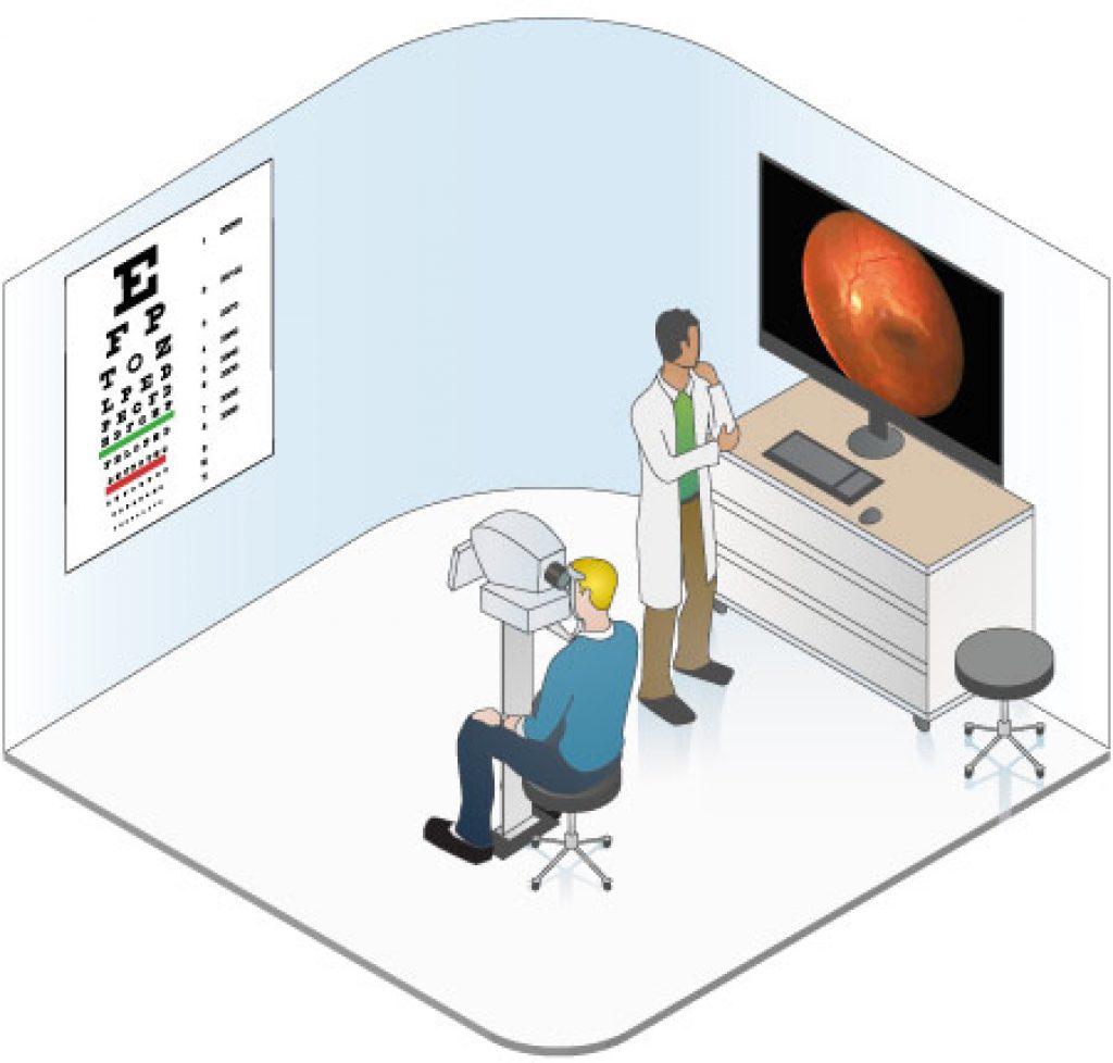
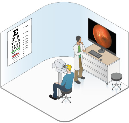
The first floor is home to our eye exam clinic. This field has grown in importance in recent years, as early detection of diseases such as diabetic retinopathy or macular degeneration can vastly increase the chances of successful treatment. The modern ophthalmologist has a variety of diagnostic devices and methods at his or her disposal.
One widely used examination device is the slit lamp microscope (or slit lamp for short). The name refers to the slit-shaped beam of light that is used to illuminate the eye, enabling further examination with a reflected light microscope. Most modern slit lamps include integrated digital cameras to document the diagnosis in photos or videos.
Physicians use a fundus camera to create high-resolution photos of the back of the eye (or 'fundus'). The newer, portable models are built to be very light and ultra-compact — which means that an embedded camera is a must. The increasing mobility of the portable devices is a significant aid to doctors and patients, as diagnosis can now be performed during house calls.
2nd Floor: Dermatology
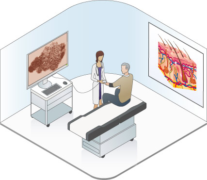
Dermatology is focused on skin diseases. Early diagnosis of diseases such as skin cancer (melanomas) can make a huge difference in successful treatment.
One common diagnostic procedure is dermatoscopy, which examines and documents suspicious skin changes using an integrated camera. This is performed over various points in time so that the development of the pigment can be recorded digitally. Special software is used not just to archive the images, but to analyze them as well. The most important benefit is the computer-supported analysis of the images integrated into the software. One key requirement for the evaluation of the recorded images is a standardized setup for capturing the images. Embedded cameras play a key role in this, compensating for different photographic conditions, including light and angle, to ensure that the images are comparable across an extended period. The color fidelity of the camera is also an important factor in keeping the diagnosis as certain as possible.
3rd Floor: Lab devices
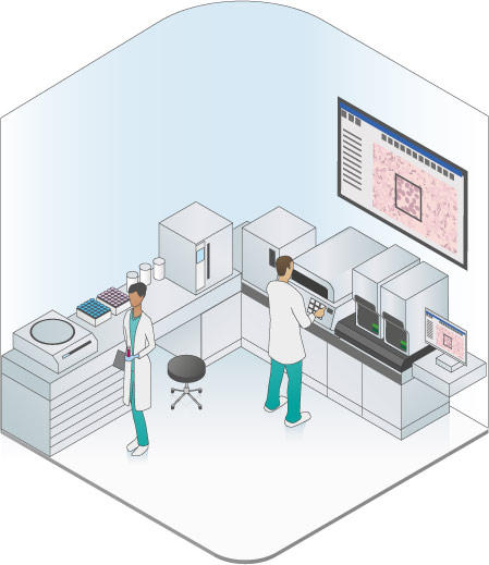
The third floor of our medical center features a dense collection of lab equipment. Many of these unremarkable-looking devices actually contain cutting-edge cameras that play a key role in the measurement process.
Fluorescent measurement is one key application performed by these tools. It involves counting the volume of proteins or other components of a substance. The more fluorescent light the camera detects, the greater the protein levels in the specimen. Samples with lower protein levels in particular require high sensitivity from the camera sensor. When properly calibrated, the cameras can even enable precise determination of values.
When a tumor is discovered in a patient, it must first be characterized before an effective therapy strategy can be designed. The assessment of the tumor tissue is an important part of this. In the past, a tissue cross-section would be examined under the microscope by a pathologist. Based on the findings, the physician would then decide whether the cells appeared to be healthy or pathological. Digital slide scanning has opened up an entirely new manner of examination. The tissue cross-section is scanned using an integrated high-resolution camera. The pathologist no longer works at a microscope, but rather reviews high-resolution images on a computer. The benefits are clear: The physician can now add comments to suspicious areas and measure or segment specific points on the image. Computer-aided diagnostics can also be used to detect pathogenic structures.
4th Floor: Operating theaters
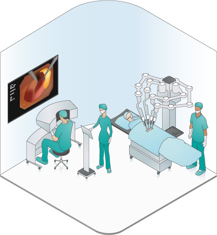
The fourth floor of the Basler Medical Center contains the operating theaters.
To maintain an overview, the operating unit's control center contains monitors that allow an observer to track the activity in all theaters at any time from a variety of angles. The cameras delivering those images are typically highly compact devices on swiveling arms placed above the operating tables. Operations on patients can then be transmitted live and/or recorded for training or consultation purposes. Devices with embedded cameras in the operating field must be high resolution and also compatible with standard hospital disinfection methods.
Over the past decade, techniques such as minimally invasive surgery have revolutionized how and when doctors decide to operate. Instead of making a large incision in the patient's abdomen, the surgeon can now access the patient's organs via a keyhole-sized opening. For orientation within the body, tiny embedded cameras built into endoscopes provide live images. In most cases, the endoscope is paired with the necessary tool, for example allowing the surgeon to constantly have the scalpel in the focus of the video. It is easy to comprehend why these cameras must be extremely reliable to ensure patient safety at all times. Endoscopic cameras typically feature highly curved lenses to provide the greatest possible field of view.
5th Floor: Research
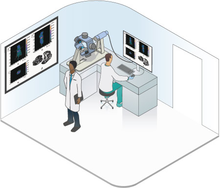
Far away from the devices involved in practical applications in daily clinical life, this topmost floor of the Basler Medical Center is reserved for research. Things are busy here, since the trend is toward ever smaller cameras for medical applications.
In recent years, researchers have developed tumor-specific antigens that are introduced into the patient's body and then adhere to the tumor. When these antigens are coupled with fluorescent markers, they can make the tumor visible. One problem is that a two-dimensional image of the tumor provides little information about its spatial structure or volume. This has led researchers to focus on bioluminescence tomography where as many as five cameras are integrated onto an O-shaped arm which, once started, rotates in a circle around the object being examined. The five cameras record images of the tumor from various viewpoints as they move. A complex set of reconstructive algorithms, not unlike those used for computer tomography (CT), then creates a 3D model of the tumor.
A view toward the future
Our Medical Center may well be growing in coming years, since cameras are expected to play an even greater role in medicine. As we've seen, embedded cameras already hold a key position in medicine and are expected to find their way into even more applications going forward.
Great things are expected from these little cameras: Beyond very strong image quality and reliability, they must also be robust and maintain their color fidelity. As the cameras grow perpetually smaller, the devices they'll be used in will also be growing more portable. This in turn opens up a new universe of potential applications, including treatment of bed-ridden patients.

