Medical Applications for Embedded Vision
Embedded vision has the potential to become the primary treatment tool in hospitals and clinics
Embedded vision and video analysis have the potential to become the primary treatment tool in hospitals and clinics, and can increase the efficiency and accuracy of radiologists and clinicians. The high quality and definition of the output from scanners and x-ray machines makes them ideal for automatic analysis, be it for tumor and anomaly detection, or for monitoring changes over a period of time in dental offices or for cancer screening. Other applications include motion analysis systems, which are being used for gait analysis for injury rehabilitation and physical therapy.
Video analytics can also be used in hospitals to monitor the medical staff, ensuring that all rules and procedures are properly followed. For example, video analytics can ensure that doctors “scrub in” properly before surgery, and that patients are visited at the proper intervals.
What are the primary vision products used in medical systems?
Medical imaging devices including CT, MRI, mammography and X-ray machines, embedded with computer vision technology and connected to medical images taken earlier in a patient’s life, will provide doctors with very powerful tools to help detect rapidly advancing diseases in a fraction of the time currently required. Computer-aided detection or computer-aided diagnosis (CAD) software is currently also being used in early-stage deployments to assist doctors in the analysis of medical images by helping to highlight potential problem areas.

Analog Devices Demonstration of the MAX78000 AI Microcontroller Performing Action Recognition
Navdeep Dhanjal, Executive Business and Product Manager for AI microcontrollers at Analog Devices, demonstrates the company’s latest edge AI and vision technologies and products at the 2024 Embedded Vision Summit. Specifically, Dhanjal demonstrates the MAX78000 AI microcontroller performing action recognition using a temporal convolutional network (TCN). Using a TCN-based model, the MAX78000 accurately recognizes a

Why Ethernet Cameras are Increasingly Used in Medical and Life Sciences Applications
This blog post was originally published at e-con Systems’ website. It is reprinted here with the permission of e-con Systems. In this blog, we will uncover the current medical and life sciences use cases in which Ethernet cameras are integral. The pace of technological transformations in medicine and life sciences is rapid. Imaging technologies used

NXP Semiconductors Demonstration of Smart Fitness with the i.MX 93 Apps Processor
Manish Bajaj, Systems Engineer at NXP Semiconductors, demonstrates the company’s latest edge AI and vision technologies and products at the 2024 Embedded Vision Summit. Specifically, Bajaj demonstrates how the i.MX 93 applications processor can run machine learning applications with an Arm Ethos U-65 microNPU to accelerate inference on two simultaneously running deep learning vision- based

Visualizing Oral Health: Various Imaging Techniques in Dentistry
This blog post was originally published at e-con Systems’ website. It is reprinted here with the permission of e-con Systems. Dental imaging has revolutionized dentistry, offering dentists a clear view beneath the surface. Explore key techniques like panoramic x-rays & intraoral cameras for improved diagnosis & treatment planning. The diagnosis and treatment of various dental

Butterfly Network’s iQ3 CMUT Sensor: The Ins and Outs
This market research report was originally published at the Yole Group’s website. It is reprinted here with the permission of the Yole Group. Innovative 9656 Transmitter CMUT MEMS die in Butterfly’s iQ3 handheld point-of-care ultrasound system. OUTLINE The iQ3, Butterfly Network’s third-generation CMUT sensor, received FDA clearance in January 2024 and follows the sale of
Major Imaging Challenges in Microscopy and How to Solve Them
This blog post was originally published at e-con Systems’ website. It is reprinted here with the permission of e-con Systems. Microscopic cameras play a major role in medical applications for surgery, pathology, and diagnostics. However, capturing clear and accurate microscopic images can be challenging due to imaging artifacts. These artifacts can appear as distortions or
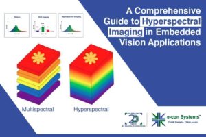
A Comprehensive Guide to Hyperspectral Imaging in Embedded Vision Applications
This blog post was originally published at e-con Systems’ website. It is reprinted here with the permission of e-con Systems. Hyperspectral imaging lets cameras see beyond human vision. It offers a high spectral resolution by capturing data in large numbers of spectral bands. Explore the technical aspects of hyperspectral imaging and discover its applications in

Vision Solutions to Enhance Your Peripheral Blood Smear Testing Devices
This blog post was originally published at e-con Systems’ website. It is reprinted here with the permission of e-con Systems. In this blog, you’ll get to learn more about this testing process and the significance of embedded camera solutions in blood smear testing. Blood smear testing has been gaining popularity for its accurate diagnosis of
Pixelation Artifact in Microscopic Imaging and Their Prevention Methods Using Advanced Camera Features
This blog post was originally published at e-con Systems’ website. It is reprinted here with the permission of e-con Systems. In microscopic imaging, pixelation is one of the most commonly occurring artifacts, as the camera subjects are micro-level. Pixelation artifact occurs when the magnification surpasses an image sensor’s capabilities, by which indistinct pixel boundaries appear.

What Causes Blooming Artifacts in Microscopic Imaging and How to Prevent Them
This blog post was originally published at e-con Systems’ website. It is reprinted here with the permission of e-con Systems. Advanced camera technologies are used with microscopes for diagnostics in the medical and life sciences industry. The occurrence of different types of imaging artifacts is a major challenge faced in microscopic imaging. Blooming or saturation

